Art, science, engineering intersect at Koch Image Gallery 2017
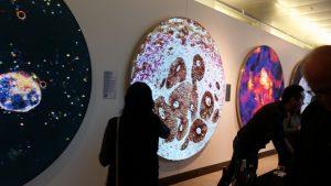
 Much enjoyed last week’s opening of the Koch Institute’s 2017 Image Awards Exhibition. The exhibit, dubbed “with/in/sight” includes 10 scientific images chosen as best-in-class from among some 120 entries from MIT life scientists and their collaborators across the country–and one from Ireland.
The display, in the public galleries at the Koch Institute for Integrative Cancer Research, is the Koch’s seventh in as many years. Its goal is to celebrate “the diversity of biomedical research at MIT and offers insight into the important role that science and engineering play in our complex and ever-changing world,” according to a Koch brochure.
The images are printed on t-shirt material stretched across frames back lit with LEDs. They are striking artwork in themselves– and showcase some of the most exciting work under way in the cancer research arena.
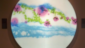
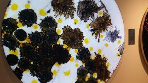
“Making Waves: Delivery for Ageless Skin.” Koch Institute, Harvard University, Mass General Hospital.
“Making Waves” conveys research on non-invasive sound waves that carry genetic material through protective layers of skin, transferring genes to cells whose genetic clocks have been turned back by the nucleic acids they have received– in order to reverse skin-aging. Credits go to Carl Schoellhammer, Denitsa Milanova, Hamberto Trevino, Cody Cleveland, Jeffrey Wyckoff, Anna Mandinova, Giovanni Traverso, Robert Langer, and George Church.
Whithead Institute: Snap Chat: A Flatworm Creates a New Profile
At the Whitehead Institute, Samuel LoCascio, Kutay Deniz Atabay and Peter Reddien are studying planarian flatworms to learn more about how they regerate. Each color in their image represents a different layer of neurons in the flatworm’s head.
Downstream Dreams: Investigating Melanoma in a Zebrafish: Koch Institute, MIT
Dahlia Perez and Jacqueline A. Lees are studying zebrafish to provide insight into melanoma. This image shows the organization of zebrafish cells in their normal state. Next, biologists will mutate a single gene known to initiate a certain melanoma in order to determine its “downstream” effects.
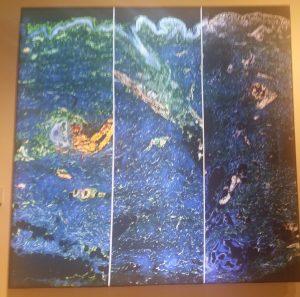
Center: “Minding the Gap: Studying the Tumor Extracellular Matrix,” Koch Institute
Tumor Penetrating Nanoparticles Infiltrate Cancer Cells, Koch Institute
Steffen RIckelt and Richard Hynes of the Koch Institute are studying not the clusters of brownish colon cancer metasteses shown in the image, screen, but, rather, the “seeming neutral” tissue matrix around them. The goal is determine how the matrix impacts the progression of tumor cells navigating a complex network of cells and proteins.
Langliang Hao, Srivatsan Raghavan, Emilia Pulver, Jeffrey Wyckoff and Sangeeta Bhatia of the Koch Institute are using biocompatible nanoparticles (yellow) to target and penetrate clusters of cancer cells (pink) with the goal of delivering treatment.
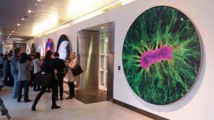
Body of Knowledge: Self-Organized Brain Cells, MIT Department of Biological Engineering and Koch Institute at MIT.
Colin Edington, Iris Lee and Linda Griffith of MIT are involved in the Griffith lab’s “Human on a Chip,” project, in which many different”mini organs”, developed from stem cells in matrix, are linked together in a bioreactor platform. The researchers are studying interactions of multiple organs and the cross between them in order to develop new disease treatments. Shown here are neurons (green) and astrocytes (red).
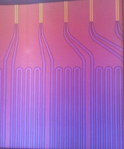
Microfluidics for the Masses, Measuring Cell Growth Rates, Koch Institute
Selim Olcum, Nathan Cermak and Scott Manalis are using microfluidics to measure the response of cell masses to drugs. Their image shows fluid filled channels (bottom) connected to tiny mass sensors shaped like hollow diving boards (top); the sensors’ whose vibrations precisely reveal the mass of individual cells passing through them. As treated cells flow across the array of sensors, each cell is weighed multiple times, thereby revealing how quickly the mass of individual cells is changing. Researchers are beginning to use this method to predict optimal treatment strategies for individual patients.
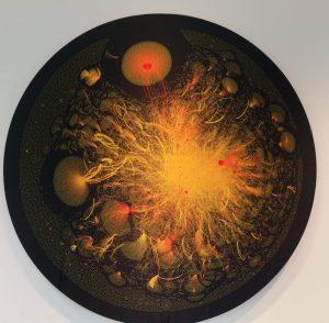
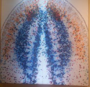
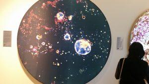
Hashtag No Filter,: Visualizing Breast Cancer Conversations. Royal COllege of Surgeons in Ireland and Wellcome Images.
My favorite image does not show cells, nor was it submitted by an MIT lab. Rather, it visualizes twitter conversations about breast cancer carried out by a network of connected cancer patients and their loved ones, patient advocates, health care professionals, and researchers. The image, by Erie Clarke, Richard Arnett and Jane Burns of the Royal College of Surgeons in Ireland, represents 92, 915 tweets posted over an eight-week period. It is from the Wellcome Images collection.
Other images not included here display pathways taken by metatastic lung cancer cells over time and ovarian cancer cells as they break through the abdominal wall.
I’m the first to admit that these photos do not do justice to the real images–nor do they adequately convey the amazing convergent technologies –including imaging–used to carry out the research.
The gallery, at street level in the Koch Institute, 500 Main Street, in Cambridge, is open to the public at no charge from 8-6 Monday-Thursday, and until 4 pm on Friday. The images are also visible from the sidewalk, outside.
Through March 2018.
Anita Harris is a writer, photographer and communications consultant based in Cambridge, MA.
New Cambridge Observer is a publication of the Harris Communications Group, an award-winning PR and market development firm located in Kendall Square, Cambridge.